- Home
- About
- Meet the Expert
- Services
- 3D Full Field Digital Mammography
- Breast Ultrasound
- Image Guided Breast Interventions
- Genetic Screening, Counselling and Testing
- Consultation and Second Opinion
- Best Breast Imaging Centre Near Me Lokhandwala, Andheri
- Best Breast Ultrasound Centre in Lokhandwala, Andheri
- Best 3D Digital Mammography in Lokhandwala, Andheri
- Best Painless 3D Mammography in Lokhandwala, Andheri |Mammocare
- Best Breast Biopsy Center in Lokhandwala, Andheri
- Best Mammography Centre in Lokhandwala, Andheri
- Blog
- FAQs
- Gallery
Cancer can be difficult to see in dense breasts on a mammogram. Ultrasound often helps to distinguish solid tumours from cysts and helps characterise lesions better.
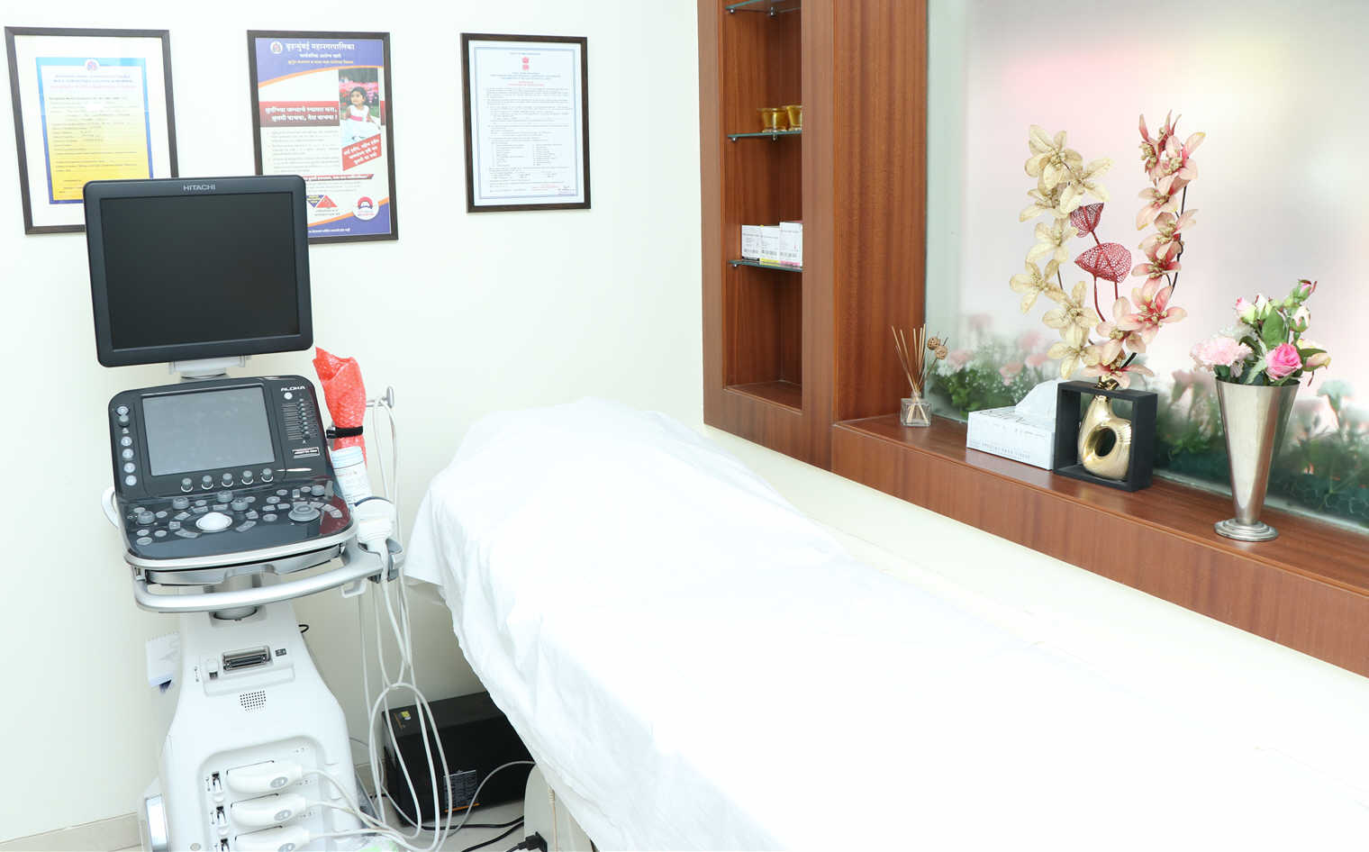
At Mammocare™, we perform a breast ultrasound in addition to mammography to help in the diagnosis. Ultrasound is safe, non-invasive and does not use radiation. We perform only ultrasound breast studies for all women below 40 years of age unless they have a breast related concern or at high risk for breast cancer. In these cases, we decide which imaging techniques should be used based on the history and clinical concerns. Ultrasound is also used for image guidance for performing breast biopsies and aspirations. Mammocare™ is equipped with one of the first new-generation ultrasound platforms from Hitachi to meet requirements of early and precise detection of the subtlest of changes on diagnostic ultrasound.

What happens during a breast ultrasound?
- You will change into a gown.
- You will lie face up, tilted slightly to the side. You will be asked to raise your arms above your head.
- The radiologist will put ultrasound gel on your skin which will feel a little cool. The radiologist will move the transducer across your skin and scan both breasts. There is usually no discomfort or pain during the examination. However, if scanning is performed over an area of tenderness, you may feel pressure or minor pain from the transducer.
- Once the imaging is complete, the clear ultrasound gel will be wiped off your skin. Any portions that are not wiped off will dry quickly. The ultrasound gel does not usually stain or discolour clothing.
Refer to our FAQ section for further information.
