- Home
- About
- Meet the Expert
- Services
- 3D Full Field Digital Mammography
- Breast Ultrasound
- Image Guided Breast Interventions
- Genetic Screening, Counselling and Testing
- Consultation and Second Opinion
- Best Breast Imaging Centre Near Me Lokhandwala, Andheri
- Best Breast Ultrasound Centre in Lokhandwala, Andheri
- Best 3D Digital Mammography in Lokhandwala, Andheri
- Best Painless 3D Mammography in Lokhandwala, Andheri |Mammocare
- Best Breast Biopsy Center in Lokhandwala, Andheri
- Best Mammography Centre in Lokhandwala, Andheri
- Blog
- FAQs
- Gallery
A biopsy is often needed when an abnormality is detected on a mammogram, ultrasound or breast MRI. These biopsies are performed based on which modality depicts the abnormality better. At Mammocare™, we offer the below-listed breast interventions. All breast interventions are performed keeping your safety as our priority.
Ultrasound-guided fine needle aspirations of lymph nodes and breast lesions
An ultrasound guided fine needle aspiration cytology test involves taking a sample from the breast with a small 22 and 24 G needles under local anesthesia. It is done to test an abnormal area without requiring surgery. Ultrasound is used to help find the area that needs to be sampled. Slides of these samples are obtained and sent to a pathology lab for testing. The entire process may take around 15minutes. You will be in the clinic for about half an hour. For breast lesions, we recommend ultrasound guided core biopsies as is done worldwide to avoid undersampling. FNACs are usually performed for axillary lymph nodes.
Ultrasound-guided core biopsies of the breast
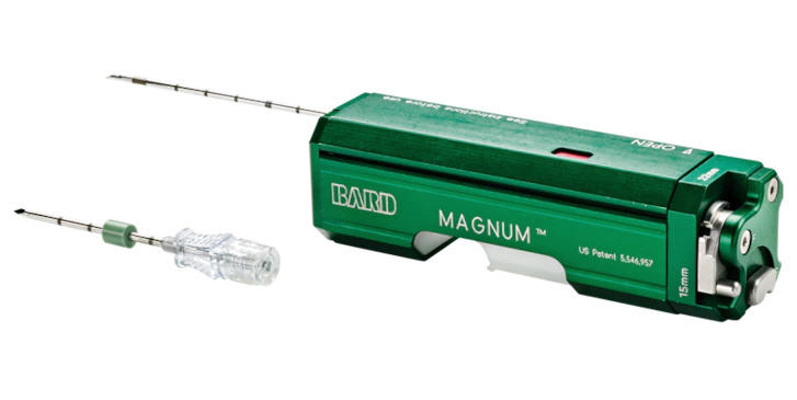
A core biopsy involves taking small samples of tissue from the breast with a special needle called a core needle. It is done to test an area of the breast tissue without requiring surgery. Ultrasound guidance is used to help find the area that needs to be sampled. The entire procedure is performed within few minutes under aseptic precautions and under local anesthesia. These samples are then sent to a pathology lab for testing. The entire process may take around 60 minutes, but it takes only 20 minutes to take the tissue samples. You will be in the clinic for about an hour. The technologist will teach you how to care for yourself after the biopsy. You may have some bruising or tenderness in your breast for a few days. This is expected and will slowly go away. At Mammocare™, we perform our biopsies using 14G needles to ensure adequate sampling.
Ultrasound guided clip/marker placement
Ultrasound guided clip placement is performed for patients with Locally Advanced Breast Cancer who have been recommended neoadjuvant chemotherapy prior to breast conserving surgery. The procedure is similar to an Ultrasound guided core biopsy and can be performed in the same sitting following the biopsy or at a later time prior to commencing neoadjuvant chemotherapy. The entire procedure is performed within few minutes under aseptic precautions and under local anesthesia. An MRI compatible metallic marker is placed within the malignant mass under ultrasound guidance. This will take only a few minutes. After this, a post procedure mammogram will be performed to ensure the position of the deployed marker. You may have some bruising or tenderness in your breast for a few days. This is expected and will slowly go away.
Ultrasound-guided aspirations of breast cysts and abscesses
Ultrasound guided needle aspirations of symptomatic breast cysts are performed at our facility under local anesthesia. If a patient presents with an inflamed and tender mass suggestive of an abscess, ultrasound guided aspiration of the pus is performed from the abnormal area within minutes under local anesthesia. The pus aspirated is then sent for culture and sensitivity analysis to diagnose the cause of infection. The entire process may take around 15minutes. You will be in the clinic for about half an hour.
Ultrasound-guided hook-wire needle localisations
When a lumpectomy is planned, the surgeon needs to be able to remove the cancer with a small amount of surrounding tissue. If the cancer is not palpable (felt be hand), image guidance is needed to place a wire within the abnormal area in the breast to guide the surgeon during surgery. Placing a wire in the abnormal area in the breast guides the surgeon where to go to find the cancer. Certain abnormalities are seen easily on ultrasound. In these cases, a short procedure is performed prior to the surgery. A hook wire is placed in the abnormal area using ultrasound guidance. At Mammocare™, we perform such procedures under sterile precautions using best techniques under local anesthesia to make it quicker and as pain free as possible.
Mammography-guided hook-wire needle localisations
When a lumpectomy is planned, the surgeon needs to be able to remove the cancer with a small amount of surrounding tissue. If the cancer is not palpable (felt be hand), image guidance is needed to place a wire within the abnormal area in the breast to guide the surgeon during surgery. Placing a wire in the abnormal area in the breast guides the surgeon where to go to find the cancer. Certain abnormalities such as microcalcifications are seen only on the mammogram and not on ultrasound. In these cases, a short procedure is performed prior to the surgery using the mammography machine. At Mammocare™, we perform such procedures under sterile precautions using best techniques to make it quicker and as pain free as possible.
Mammography-guided stereotactic vacuum-assisted biopsies [coming soon]
A core biopsy involves taking small samples of tissue from the breast with a special needle called a core Biopsy needle. It is done to test an area of the breast tissue without surgery. Certain abnormalities are seen only on the mammogram which are not seen on ultrasound. In these cases, a mammography guided stereotactic biopsy is advised. During such a stereotactic biopsy, a special mammography machine is used. It will help the radiologist find the area that needs to be sampled. These samples are then sent to a pathology lab for testing.
Please download the care instructions if you have been scheduled for a biopsy. If you have been advised a biopsy following your mammogram or ultrasound at another facility/center, feel free to walk in at our clinic with your reports and films if you have any queries. Unless you need to be off certain medications, biopsies and breast interventions are performed on the same day at Mammocare™.

For more information, see:
